In the second half of the XVII century, the Dutch scientist Antoni Van Leeuwenhoek described the wonderful worlds revealed by his amazing microscopium in hundreds of letters sent to the Royal Society of London. In this correspondence he told of the first microscopic unicellular organisms massed in a single drop of stagnant water, correctly calculating that it could host a number many times larger than the sum of humans on the Earth, he told of spermatozoa, which he defined as animalculi (small animals), he wrote of blood cells, capillaries and muscle fibers. In 1683, he pointed his microscope at the teeth of two elderly volunteers and described the first microscopic observation of bacteria, ten times smaller than a eukaryotic cell, something that no one else would have been able to replicate for more than a century.
These observations, apparently too successful to be true, aroused great skepticism in contemporary scientists, who in the same years pointed their frosted- or blown-glass lenses to see the invisible, but received nothing in return comparable to what van Leeuwenhoek described. Among the most eager to know the secrets of those lenses was Robert Hooke, who in 1665 had first observed the microscopic cells of a cork bark, thereby coining the term cellula (Latin for “small room”, to which those basic units resembled). Hooke had summarized his observations under the microscope in a very popular scientific treatise, Micrographia (1665), accompanied by fifty-eight engravings including the famous one of a flea.
In the vane attempt to persuade Van Leeuwenhoek to reveal his secrets, Hooke had published instructions for producing blown-glass spherical lenses for use in microscopes already in 1665, and again in 1678. But in 1711 Van Leeuwenhoek, then 80 years old, led a group of German aristocrats to believe that he had discovered a new method of working glass that “allowed the production of non-spherical lenses”, implying that this was the secret of the magnification power of his microscopes. However, a recent neutron tomography study carried out on the very glasses of Leeuwenhoek’s microscopes [T. Cocquyt et al., Neutron tomography of Van Leeuwenhoek’s microscopes. Science Adv. 7, 2402 (2021)] revealed that there was no secret at all but his ability: his lenses were ordinary spherical segments, only carved with exceptional precision from a carefully blown glass without any air bubbles trapped in.
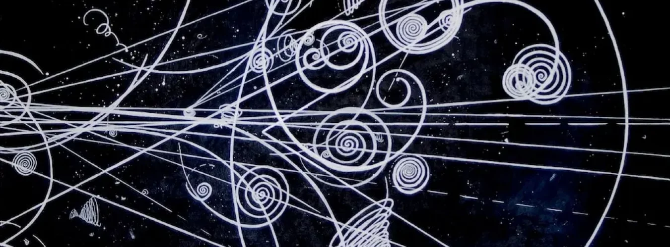
In a letter of September 7, 1674, Van Leeuwenhoek writes: About two leagues from this town there lyes an inland sea called Berkelsee–meer. (…) Observing the water (…) I took up some of it in a glass-vessel, which having viewed the next day, I found moving in it several earthy particles, and some green streaks, spirally ranged, (…) among all of which there crawled abundance of little animals, some of which were roundish; those that were somewhat bigger than others were of an oval figure: ln these latter I saw two legs near the head and two little fins on the other end of their body. (…) The motion of most of them in the water was so swift, and so various, upwards, downwards, and round about, that I confess I could not but wonder at it. Oh, that one could ever depict so wonderful a motion! I judge, that some of these little creatures were above a thousand times smaller than the smallest ones, which I have hitherto seen. From the description of shapes and colors and heads and fins, the renowned biologist and historian of science Clifford Dobell concluded that Leeuwenhoek saw rotifers, ciliates, and a common species of Euglena.
The generation of forces and the ability to move represent some of the most striking abilities of living cells. Prominent examples are indeed cell motility, as well as the contraction of muscles, but also active transport of materials and of organelles inside a cell, for example during cell division and mitosis. Such movements are generated at the molecular level by protein molecules, specialized to work by interacting with filaments of the cell inner supporting structure, the cytoskeleton. Such molecular-scale motors consume adenosin-triphosphate (ATP) as a fuel, and convert its chemical energy to mechanical work. Myosin motors, for example, can generate motion along the filaments of actin, that is those linear supporting proteins which run everywhere inside a cell like a spiderweb. Kinesin and dynein motors move instead along microtubules, the thicker filaments of the cellular structure. The motion of such molecules must evidently be subject to thermodynamical fluctuations, which at the scale of the single molecule can completely hinder the kinetics of motion. To overcome this Brownian noise, the molecular motors use mechanisms of thermal ratcheting, which can break the left-right symmetry of the stochastic displacement, and make the motor to move in a well-defined direction. Among many examples available, you may have a look at this video showing myosin molecules running on immobilized actin filaments in vitro, at the trailblazing speed of about 1 meter per week (https://www.youtube.com/watch?v=avm_d2CTiqw), which is however enough to traverse a cell diameter in a few seconds.
Notably, in certain situations cells can generate oscillatory motion and operate as mechanical oscillators at the very molecular scale (not to be confused, e.g., with protein expression periodicity, or the larger scale rhythms of the cell cycle). Oscillatory behaviors of muscle cells have been known for a long time, e.g. in the case of some flight muscles which move the wings of insects. Many insects such as wasps and bees have muscles called ‘asynchronous’, which generate oscillatory contractions by a mechanism located in the muscle itself, which drives the beating of the wings. For such muscles, each single impulse coming from the motor neurons can elicit several (5 to 10) muscle impulses in rapid succession. This type of muscle differs from the ‘synchronous’ flight muscles found in vertebrate birds, in which contractions are triggered by each periodic nerve signal in one-to-one phase with the muscle contraction cycle, and are not independently generated in the muscle. More recently, oscillatory behaviors have been observed under certain conditions in fibrils of ordinary skeletal muscles, which under normal conditions would not oscillate.
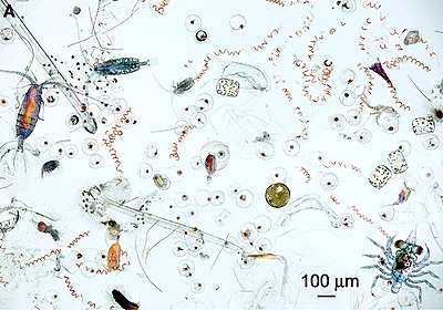
The euglenids observed by Van Leeuwenhoek among many other animalculi, are unicellular eukaryotes that situate somewhere between animals and plants. They are believed to have originated from a primitive unicellular organism that may have swallowed (“endocytosed”) a green unicellular alga. Such cells lack a real cytoskeleton, but rather have a homogeneously surface-distributed protein network capable of deforming the cell membrane. These organisms have developed a unique low-Reynolds number swimming technique, still quite poorly understood, based on an oscillatory recurrent deformation of the cell body membrane. The power stroke of an euglenid consists of a large distortion of the membrane that starts at the head and propagates back along its slender body. The overall shape of the membrane looks like if a sphere is sliding down a thin coaxial cylinder, opposite to the euglenid’s direction of motion. You may think of a spherical bead sliding on a necklace, as the necklace is being pulled through the water. Once the large spheroid distortion reaches the end, the body of the euglenid is rearranged, and the next stroke is initiated, in a periodic motion of typical frequency of one stroke per second. This singular motion pushes forward the cell body at the expense of the volume of water displaced, and results in a swimming velocity of the order of 40 microns/second. Considering that the euglenid is about 40-50 microns in length, its comparative speed (body-lengths/second) is twice that of a blue dolphin, which swims at peak speeds of 15 km/h and measures 6-8 m head to tail. The minimum force necessary to maintain the euglenid speed can be estimated by the Stokes’ drag formula for the spherical part of the body, which definitely presents the largest resistance to the fluid. By calculating F=6phRv, with h=1 mPa per second the water viscosity, and R~5 microns, we get F~4 pN (piconewton). The expended power is obtained as P=Fv~1,6 × 10⁻¹⁶ W. Since a mole of ATP gives off 30.5 kJ of energy, or 5.07 × 10⁻²⁰ J per molecule, the euglenid must use about 3,000 ATP molecules per second to maintain its swimming speed. Notably, this figure is 10 to 100 times larger than the value per unit mass compared to an athlete swimming breaststroke, showing that the water viscosity has a totally different effect at the micron scale compared to the macroscopic scale: for euglena, water rather feels like molasses.
In a paper just appeared in Nature Nanotechnology (Probing nanomotion of single bacteria with graphene drums, April 2022) by the group of Cees Dekker in Delft, the vibrational movement of Escherichia coli bacteria was detected with a novel single-cell technique based on suspended graphene “drums”. Such devices are made of ultrathin (<1 nm) bilayer CVD graphene that covers circular cavities etched in a SiO2 matrix, with a diameter of 8 µm and a depth of 285 nm. A chip comprising an array of many thousands of these graphene-covered cavities was placed inside a cuvette containing E. coli bacteria in a proper cell culture medium, mixed with a chemical to bind the bacteria to the graphene surface. The nanomotion of a bacterium resulted in a time-dependent deflection z(t) at the center of the suspended membrane, which was measured using laser interferometry from the modulation of the intensity of the reflected light. The small mass, high stiffness, and micron-sized area of the suspended graphene “drum” enables detecting nanomotion even at the single bacterium level. The vibrations produced by different E. coli strains allowed to investigate the contributions of the different parts of the bacterium body, demonstrating that the rotating movement of the flagellum provides the main source of nanomotion. Moreover, by tracing the nanomotion in the presence of antibiotics, they could show that this novel ultrasensitive graphene-based platform may enable antibiotic susceptibility tests with single-bacterium sensitivity.
The results of their vibrational analysis are consistent with those found for bacterial colonies on AFM cantilevers, and show that the nanomotion generated by even a single E. coli bacterium, does not follow a specific periodicity, but instead involves a wide range of frequencies, with a possibly fractal power spectrum. To make the story funnier, however, the drum vibration induced by the bacterium can also be clearly perceived by listening to audio recordings, which were generated by converting the interferometric traces to a sound track. In the words of Cees Dekker, “What we saw and heard was striking! When a single bacterium adheres to the surface of a graphene drum, it generates random oscillations with amplitudes as low as a few nanometers, that we could detect. We could hear the sound of a single bacterium!” Single bacteria were observed to produce peak fluctuations of up to 60 nm in amplitude, corresponding to forces of up to 6 nN. These forces are larger than the typical pN-scale forces generated by a single molecular motor, and even larger than flagellar motors, of the order of ~100 pN, but are similar to the forces measured by AFM spectroscopy on membranes of single Saccharomyces cerevisiae cells.
“A rock band is only as good as its drummer” it was said, and examples abound. The Beatles skyrocketed to fame after they replaced Pete Best with Ringo Starr; R.E.M. music went lost in a vanilla patchiness after Bill Berry retired to a cow farm; Led Zeppelin disbanded after John Bonham died in 1980; Keith Moon’s death did not stop The Who, but you rather wish it had… ; the Clash fired Topper Headon in 1982, and later admitted it was the beginning of their end; Keith Richards repeatedly said the only event that would force Rolling Stones to retirement would be the departure of Charlie Watts… well, that is history already, and the Stones are still playing, we’ll see for how long still. Anyway, with E.coli beating the tempo on graphene drums we got our nano-drummer. To build the first rock nanoband we must now find just a nanoguitar and a nanobass.